What is melanoma?
Melanoma Symtoms
Symptoms of melanoma include:
- A large brownish or black spot with darker speckles
- A mole that changes in color, size or feel or that bleeds
- A small lesion with an irregular border and portions that appear red, white, blue or blue-black
- Dark lesions on your palms, soles, fingertips or toes, or on mucous membranes lining your mouth, nose, vagina or anus
Malignant melanoma can metastasize to other organs and can lead to metastatic melanoma and death.
Where does melanoma develop on the body?
Melanoma can develop anywhere on your body. They may develop in an existing mole in 20 – 30 % of individuals or in otherwise normal skin.
Men Bodies
In men, melanoma most often appears on the face or the trunk.
Women Bodies
In women, this type of cancer most often develops on the lower legs. In both men and women, melanoma can occur on skin that hasn’t been exposed to the sun.
Skin Tone and Melanoma Appearance
Melanoma can affect people of any skin tone. In people with darker skin tones, melanoma tends to occur on the palms or soles, or under the fingernails or toenails.
Malignant melanoma warning signs:
The following are warning signs of having a melanoma and melanoma pictures:

Types of Malignant Melanoma
There are four types of melanoma. Three of them begin as melanoma in situ. This means they start in the top layer of the skin. They then can become more invasive with time. The fourth is invasive from the start. Invasive melanomas are more serious. They penetrate deeper into the skin and may spread to other areas of the body.
1. Superficial spreading melanoma
This is the most common type. They account for 70 percent of melanomas. This is most often seen in young people. As the name suggests, this melanoma grows along the top layer of the skin for some time before penetrating more deeply.
The first sign is the appearance of a flat or slightly raised discolored patch that has irregular borders and is somewhat asymmetrical in form. The color varies, and you may see areas of tan, brown, black, red, blue or white. This type of melanoma can occur in a previously benign mole or arise as a new lesion.
It can be found almost anywhere on the body but is most likely to occur on the trunk in men, the legs in women, and the upper back in both.
2. Lentigo maligna
This melanoma is similar to the superficial spreading type.
It too remains close to the skin surface for a while, and usually appears as a flat or mildly elevated, mottled, tan, brown or dark brown discoloration. This type of in situ melanoma is found most often in the elderly.
It arises on chronically sun-exposed, damaged skin on the face, ears, arms and upper trunk. Lentigo maligna is the most common form of melanoma in Hawaii. When this cancer becomes invasive, it is referred to as lentigo maligna melanoma.
3. Acral lentiginous melanoma:
This melanoma also spreads superficially before penetrating more deeply.
It is quite different from the others, though, as it usually appears as a black or brown discoloration under the nails or on the soles of the feet or palms of the hands. This type of melanoma is sometimes found on dark-skinned people and tends to advance more often than superficial spreading melanoma and lentigo maligna because it is detected later.
It is the most common melanoma in African-Americans and Asians.
It is the least common melanoma in Caucasians.
4. Nodular melanoma
This melanoma is usually invasive at the time it is first diagnosed.
The malignancy is recognized when it becomes a bump. It is usually black, but occasionally is blue, gray, white, brown, tan, red or skin tone.
The most frequent locations are the scalp, trunk, legs and arms. It is found mainly in elderly people. This is the most aggressive and deadly of the four types of melanoma.
There is a variant of melanoma which has no pigment and can be whitish in color. It is known as an amelanotic melanoma. Since they do not have pigment, they can go unnoticed. There can be a delay in diagnosis and thus a poorer prognosis.
What does melanoma look like?
Melanoma Pictures Melanoma Photos Malanoma Cancer Pictures

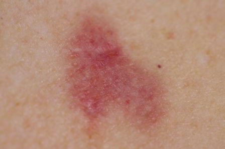


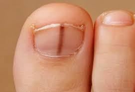


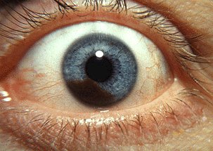
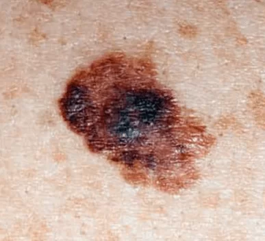

What are the stages of melanoma?
- Stage 0 Melanoma (Melanoma in situ): Cancer is confined to the epidermis (outer skin layer). No spread to lymph nodes or distant sites. Highly treatable, often cured with surgical removal.
- Stage 1 Melanoma: Thin melanoma, less than 2mm thick, no ulceration (IA) or with ulceration (IB). No lymph node involvement or distant spread. High survival rate with surgery.
- Stage 2 Melanoma: Thicker melanoma (>2mm), may be ulcerated. No lymph node or distant spread. Subdivided into IIA, IIB, IIC based on thickness and ulceration. Surgery is primary treatment, but risk of recurrence increases.
- Stage 3 Melanoma: Cancer has spread to nearby lymph nodes or skin (regional spread) but not distant organs. Subdivided into IIIA, IIIB, IIIC, IIID based on tumor thickness, ulceration, and extent of lymph node involvement. Treatment may include surgery, immunotherapy, or targeted therapy.
- Stage 4 Melanoma: Melanoma has metastasized to distant lymph nodes, organs (e.g., lungs, liver, brain), or other areas. Most serious stage, requiring aggressive treatments like immunotherapy, targeted therapy, or clinical trials. Prognosis is poorer but varies.
What is the treatment for melanoma?
- Stage 0 (Melanoma in situ):
- Surgery: Wide local excision to remove the melanoma and a small margin of healthy skin. Cure rate is nearly 100% if completely removed.
- Stage I and II:
- Surgery: Wide local excision with a larger margin (1–2 cm) of surrounding skin. Sentinel lymph node biopsy may be performed for thicker tumors to check for spread.
- Adjuvant Therapy (for high-risk Stage II): Immunotherapy (e.g., pembrolizumab or nivolumab) or targeted therapy (e.g., dabrafenib and trametinib for BRAF-mutated melanoma) may be used to reduce recurrence risk.
- Stage III:
- Surgery: Excision of the primary tumor and affected lymph nodes (lymphadenectomy).
- Immunotherapy: Drugs like pembrolizumab, nivolumab, or ipilimumab to boost the immune system’s ability to fight cancer.
- Targeted Therapy: For BRAF-mutated melanomas, drugs like vemurafenib, dabrafenib (BRAF inhibitors), combined with MEK inhibitors (e.g., trametinib, cobimetinib).
- Radiation Therapy: Rarely, to control symptoms or local recurrence in specific areas.
- Stage IV:
- Immunotherapy: Checkpoint inhibitors (e.g., pembrolizumab, nivolumab, or nivolumab plus ipilimumab) to enhance immune response against cancer cells.
- Targeted Therapy: For BRAF mutations (present in ~40–50% of melanomas), combinations like dabrafenib + trametinib or encorafenib + binimetinib.
- Chemotherapy: Less common but may use drugs like dacarbazine if other treatments aren’t suitable.
- Radiation Therapy: To relieve symptoms in areas like the brain or bones.
- Surgery: For isolated metastases (e.g., in the lung or brain) to remove tumors in select cases.
- Clinical Trials: Novel therapies, such as new immunotherapies or combination treatments, for advanced cases.
- General and Supportive Treatments:
- Palliative Care: To manage symptoms and improve quality of life, especially in advanced stages.
- Skin Monitoring: Regular follow-ups with dermatologists to detect recurrence or new melanomas.
- Lifestyle Adjustments: UV protection to prevent further skin damage.
- Early-stage melanomas (0–I) are often cured with surgery alone. Advanced stages (III–IV) require a combination of therapies, with outcomes varying based on response.
- Immunotherapy and targeted therapies have significantly improved survival rates for advanced melanoma Richmond VA since the 2010s.
- Treatment plans are personalized, often guided by genetic testing (e.g., BRAF mutation status) and multidisciplinary teams.
- Side effects vary: surgery may cause scarring; immunotherapy can cause fatigue, skin reactions, or autoimmune issues; targeted therapies may cause fever or joint pain.
Is melanoma curable?
- Stage of Melanoma: Melanoma is staged from 0 (in situ, confined to the top skin layer) to IV (advanced, spread to distant organs).
- Early stages (0–II): Highly curable with surgery alone in most cases.
- Later stages (III–IV): More challenging, but treatments like immunotherapy and targeted therapies have improved outcomes significantly in recent years.
- Survival Rates: These are estimates based on large-scale data (e.g., SEER database from NCI). They represent 5-year relative survival rates (chance of surviving compared to the general population):Note: These rates have improved over time due to better treatments; for example, drugs like pembrolizumab (Keytruda) and ipilimumab (Yervoy) have boosted survival in advanced cases.StageLocalized (no spread)Regional (spread to nearby lymph nodes)Distant (metastasized)Overall5-Year Survival Rate~99%~68%~30%~94% (all stages combined)
- Surgery: The primary cure for early melanoma. Wide local excision removes the tumor and surrounding tissue. Cure rates exceed 90–95% for thin melanomas (<1 mm thick).
- Immunotherapy: Boosts the immune system to fight cancer (e.g., checkpoint inhibitors like nivolumab). Effective for stage III–IV; can lead to long-term remission in 40–50% of advanced cases.
- Targeted Therapy: For melanomas with BRAF gene mutations (about 50% of cases), drugs like dabrafenib + trametinib shrink tumors and extend survival.
- Radiation, Chemotherapy, or Clinical Trials: Used less often now, as they’re less effective than newer options. Chemotherapy has response rates of only 10–20% in advanced melanoma.
- Recurrence Risk: Even if “cured,” follow-up is crucial, as melanoma can return. Regular skin checks and imaging help catch it early.
- Avoid excessive sun exposure, use SPF 30+ sunscreen, and skip tanning beds (which increase risk by 75% per ACS data).
- Self-exams: Look for ABCDE signs (Asymmetry, Border irregularity, Color variation, Diameter >6mm, Evolving changes).
- Screening: Annual dermatologist visits for high-risk individuals (fair skin, many moles, family history).
Is melanoma deadly?
- Aggressive Nature: Unlike basal or squamous cell skin cancers (which rarely spread and have near-100% survival rates), melanoma arises from melanocytes (pigment-producing cells) and can invade deeply or metastasize via blood or lymph systems. About 20–30% of melanomas are diagnosed at an advanced stage in some populations.
- Risk Factors Increasing Deadliness:
- Thick tumors (>4 mm deep) or those with ulceration.
- Genetic mutations (e.g., BRAF in ~50% of cases).
- Weakened immune system, older age, or male sex (men have higher mortality rates post-diagnosis).
- Delay in diagnosis: Metastatic melanoma historically had a median survival of 6–9 months before modern therapies.
- Global Impact: The WHO estimates ~325,000 new melanoma cases and ~57,000 deaths worldwide in 2020 (latest comprehensive data). In the U.S., the ACS projects about 8,300 deaths from melanoma in 2024 out of ~100,000 new cases—making it responsible for ~1% of all skin cancer diagnoses but ~75% of skin cancer deaths.
Stage at Diagnosis | Description | 5-Year Survival Rate | Approximate % of Cases Diagnosed at This Stage |
|---|---|---|---|
Localized (Stage 0–II) | Confined to skin; no spread | 99% | ~83% |
Regional (Stage III) | Spread to nearby lymph nodes | 68% | ~13% |
Distant (Stage IV) | Metastasized to distant organs | 30% | ~4% |
Overall (All Stages) | – | 94% | – |
- Mortality Trends: Death rates have dropped ~5% per year since 2015 in the U.S., per ACS, due to better screening and drugs. For stage IV, median survival is now 2–3 years or more with treatment (vs. months pre-2011).
- Lifetime Risk: About 1 in 38 Americans will develop invasive melanoma; risk of dying from it is ~1 in 200 if screened regularly.
- Early Detection: Thin melanomas (<1 mm) have a 95–99% cure rate with surgery alone. Regular self-exams (ABCDE rule: Asymmetry, Border, Color, Diameter, Evolving) and dermatologist visits can catch 90% early.
- Treatments for Advanced Cases:
- Surgery, immunotherapy, or targeted therapy can extend life significantly; some patients achieve “no evidence of disease” status.
- Clinical trials (e.g., via ClinicalTrials.gov) offer options like TIL therapy or vaccines.
- Prevention: UV exposure causes 90% of melanomas. Use broad-spectrum sunscreen (SPF 30+), avoid peak sun hours (10 a.m.–4 p.m.), and never use tanning beds—reducing risk by up to 50%.
Does melanoma itch?
How long does it take melanomas to spread?
- Tumor Thickness and Type:
- Thin melanomas (<1 mm deep): Often indolent (slow-growing); may take 5–10+ years to spread, if at all. These account for ~70% of diagnoses and rarely metastasize quickly.
- Thicker ones (>4 mm) or ulcerated: Faster progression; can spread in 6–18 months.
- Subtypes:
- Nodular melanoma (15–20% of cases): Most aggressive; vertical growth phase can lead to spread in 3–12 months.
- Superficial spreading (70%): Slower; horizontal phase lasts 1–5 years before deepening and potentially spreading.
- Lentigo maligna: Very slow; often 10–20 years in sun-damaged skin of older adults.
- Acral or mucosal: Variable but can be rapid due to delayed detection.
- Mitotic Rate and Ulceration: High cell division (mitoses >1 per mm²) or broken skin surface accelerates spread by 2–5x, per AJCC staging.
- Genetics and Host Factors: BRAF mutations speed growth; stronger immunity (e.g., via T-cells) can delay it. Men, older age (>60), and immunosuppression (e.g., from organ transplants) increase risk of quicker metastasis.
- Location: On the head/neck or trunk: Slightly faster spread potential than limbs.
- Radial (surface) growth phase: Months to years (no metastasis yet).
- Vertical invasion and metastasis: Can occur after 1–2 years in average cases, but data shows 20–30% of melanomas metastasize within 5 years of diagnosis if untreated.
Stage | Description | Time to Reach This Stage (from onset) | Risk of Further Spread | 5-Year Survival |
|---|---|---|---|---|
0 (In Situ) | Confined to epidermis; no invasion | Years (often 2–10+; precancerous phase) | Very low (<5%) | ~99% |
I–II (Localized) | Invades dermis but not beyond skin | 1–5 years | Low (5–20% metastasize in 5–10 years if thin) | 90–99% |
III (Regional) | Spread to nearby lymph nodes | 1–3 years post-invasion | Moderate (recurrence in 20–50% within 2 years) | 40–78% |
IV (Distant) | Metastasized to organs (lungs, liver, brain) | 6 months–2 years after regional spread | High (rapid progression; median survival 6–24 months pre-modern treatments) | 15–30% |
- Key Insight: Spread isn’t linear. Doubling time for melanoma cells can be 1–3 months in aggressive cases (per imaging studies). Once in lymph nodes, distant metastasis can follow in weeks to months via blood vessels.
- Statistics on Speed: Per a 2020 Journal of Clinical Oncology review, ~4% of cases are metastatic at diagnosis (missed early phase). For stage II, 10-year metastasis risk is 10–30%; for stage III, half recur within 2 years.
How quickly should melanoma be removed?
Can basal cell carcinoma turn into melanoma?
Can melanoma appear overnight?
- Biological Timeline: Melanoma starts with atypical melanocytes (pigment cells) multiplying abnormally. This radial growth phase (surface spreading) can take months to years before becoming visible or raised. Vertical invasion (deepening into skin layers) follows, potentially leading to symptoms. Studies in Journal of the American Academy of Dermatology show most melanomas evolve from pre-existing nevi (moles) over 6–24 months, with only ~20–30% arising de novo (on clear skin).
- Earliest detectable changes: Often microscopic; visible spots need time to reach 1–2 mm.
- Aggressive subtypes (e.g., nodular melanoma, 15–20% of cases): Can grow faster (doubling in 1–3 months) and appear as a new bump or nodule in weeks, mimicking “sudden” onset. These are dark, firm, and dome-shaped but still require prior cellular buildup.
- Perception vs. Reality: Hormonal changes (e.g., pregnancy), trauma, or inflammation can make a dormant mole darken or swell temporarily, creating an illusion of overnight appearance. Or, you might simply notice it for the first time after ignoring subtle prior signs.
- Stats on Onset: Per NCI data, the average time from cellular mutation to diagnosis is 2–5 years. Less than 5% of patients report “sudden” new lesions without prior moles, and even those had underlying changes detectable via dermoscopy (magnified skin exam).
Why consult Dr. Robert DeConti in Richmond, VA, for Melanoma Treatment?
- Specialized Expertise in Skin Cancer Surgery: Dr. DeConti is a leading specialist in Mohs micrographic surgery (and “Slow Mohs” techniques) for precise removal of skin cancers, including melanoma, basal cell, and squamous cell carcinomas. This method minimizes tissue removal while ensuring complete cancer excision, which is crucial for melanoma to prevent recurrence and achieve clear margins. He also employs advanced techniques like Complete Circumferential Peripheral and Deep Margin Assessment (CCPDMA) for thorough tumor evaluation.
- Focus on Melanoma and Related Conditions: His practice at DeConti Plastic Surgery emphasizes dermatology evaluations and treatments for melanoma, atypical moles, precancerous lesions, and tumors. He integrates laser therapies to enhance outcomes and reduce scarring, combining surgical precision with cosmetic results—ideal for visible areas affected by melanoma.
- Reconstructive Skills for Optimal Recovery: As a reconstructive plastic surgeon, Dr. DeConti excels in post-excision reconstruction, such as scar revision and keloid management, to restore appearance and function after melanoma removal. This holistic approach addresses both the medical and aesthetic aspects of treatment, which is especially important for early-stage melanomas treated surgically.
- Patient-Centered Care and High Satisfaction: Patients praise Dr. DeConti for his compassionate, attentive approach and ability to detect issues early—e.g., one review credits him with identifying and removing a potentially cancerous lesion during a routine procedure, potentially preventing advanced skin cancer. His practice is described as a “class act” with personalized follow-up, including thoughtful gestures like recovery flowers.
- Accessibility and Affiliations: Located at 7229 Forest Ave, Suite 101, Richmond, VA, his office accepts major insurances (including Medicare) and new patients. He is affiliated with top local hospitals like Henrico Doctors’ Hospital, Retreat Doctors’ Hospital, and Bon Secours facilities, ensuring seamless care for more advanced cases requiring multidisciplinary input (e.g., immunotherapy referrals).
- Proven Track Record: A University of Virginia School of Medicine graduate, Dr. DeConti has been featured on NBC, ABC, and Fox for advancements in surgical techniques. His state-of-the-art facility offers in-office procedures, reducing the need for hospital stays.